Dyslexia symptoms and PTSD- is there a connection?
How the left brain and the corpus callosum are effected by ptsd as well as dyslexia
What is dyslexia?
"Dyslexia is a specific learning disability that is neurobiological in origin. It is characterized by difficulties with accurate and fluent word recognition and by poor spelling and decoding abilities." (Reid, et. al., 2003)
Some people who had dyslexia: Albert Einstein, Pablo Picasso, Leonard Da Vinci, Thomas Edison. Articles on dyslexia
"Dyslexia occurs in children with normal vision and normal intelligence." Mayo Clinic
What are symptoms of dyslexia?
"There is no single pattern of difficulty that affects all dyslexic people." Read a list of symptoms
Is it possible to improve the brains function? Yes.
"The three main types of dyslexia are trauma dyslexia, primary dyslexia and developmental dyslexia.
- Trauma dyslexia usually occurs after some type of brain trauma or injury to the area of the brain that controls reading and writing.
Primary dyslexia is a dysfunction of, rather than damage to, the left side of the brain (cerebral cortex) and does not change with maturity. Individuals with this type are rarely able to read above a fourth grade level and may struggle with reading, spelling, and writing as adults. Primary dyslexia is hereditary and is found more often in boys than in girls.
Developmental dyslexia is caused by hormonal development during the early stages of fetal development. Developmental dyslexia diminishes as the child matures. This type is also more common in boys." Reading Success Lab
http://www.medicinenet.com/dyslexia/article.htm
"'Trauma dyslexia' usually occurs after some form of brain trauma or injury to the area of the brain that controls reading and writing."
What are some of the causes of dyslexia?
"Dyslexia seems to be caused by a malfunction in certain areas of the brain concerned with language." Mayo Clinic
"Studies show that individuals with dyslexia process information in a different area of the brain than do non-dyslexics." International Dyslexia Association "Neurological research suggests that there may be some abnormality in the function of the left side of the brain which controls the lexical system" The Dyslexia Institute
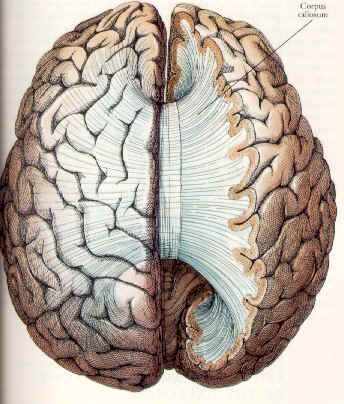
"There are thought to be various main factors within the brain that contribute to dyslexia. Two of those factors are linked an underutilized left hemisphere and a central bridge of tissue in the corpus callosum."
That is to say the:
- Damage or under-use of the left side of the brain and
- The connecting tissue between the two sides of the brain.
More about the corpus callosum and dyslexia:
"One section of the brain which is intimately involved in cerebral organization, both during growth and all through adulthood, is the corpus callosum. This thick bridge of neural tissue in the middle of the brain connects the two hemispheres, conveying information from one side to the other. "
"It seems reasonable to assume that without the fast, accurate guidance of a central control mechanism, the brain might show the kinds of symptoms which we see in dyslexia." Research on the corpus callosum and dyslexia articles on dyslexia
PTSD and the sides of the brain
"When persons with posttraumatic
stress disorder remember trauma, right areas of their brains tend to be
activated, whereas when individuals without PTSD remember trauma, left
areas of their brains are apt to be aroused, according to a study reported
in the January American Journal of Psychiatry." Psychiatry
online
"The right brain controls
the left side of the body and the left brain controls the right side of
the body. The right brain is the more creative or emotional hemisphere
and the left brain is the analytical and judgmental hemisphere. Anything
that is new or not familiar to an individual is right brain dominant.
Anything that is familiar is left brain dominant." A
very simple explanation of the brain
Sexual abuse, PTSD and the corpus callosum
"Abused children with PTSD have lower intracranial and cerebral volumes, larger lateral ventricles, and a smaller corpus callosum than healthy controls, which may indicate neuronal loss"
"In the PTSD children, we saw that the corpus callosum did not grow with age compared with controls, which may be due to a failure of myelination," said DeBellis."
Is it really possible that psychological trauma causes changes in the brain?
"It appears as if the psychological impact of childhood physical abuse can damage the corpus callosum—the major information pathway between the two brain hemispheres. In one of their studies, Teicher and his coworkers found that sexual abuse in girls was associated with a major reduction in the size of the corpus callosum. (This result was independently replicated by Michael DeBellis, M.D., a psychiatrist at the University of Pittsburgh.)" Psychiatric News
Although there have been fewer studies on adult PTSD victims there is evidence that the corpus callosum is effected in adults as well.
"Neuroimaging studies indicate that PTSD in children is associated with diffuse CNS effects (i.e., smaller cerebral volumes and corpus callosum areas)" Am J Psychiatry
Articles on dyslexia and ptsd, the corpus callosum and sexual abuse
"Teachers may use techniques involving hearing, vision and touch to improve reading skills. Helping a child use several senses to learn — for example, by listening to a taped lesson and tracing with a finger the shape of the words spoken — can help him or her process the information. The most important teaching approach may be frequent instruction by a reading specialist who uses these multisensory methods of teaching." Mayo Clinic
Other treatments for dyslexia and ptsd
Improvement in brain activity is possible See articles on treatment below.
Discussion
board about dyslexia
References and Research links:
Reid L., et. al., (2003) A Definition of Dyslexia. Annals of Dyslexia, 53, p1-14.
References- articles on dyslexia, ptsd and sexual abuse
International Dyslexia Association
Multiple victimization- Child sexual assault victims are often revictimized later in life.
Dyslexia and corpus callosum morphology
Corpus Callosum Morphology, as Measured with MRI, in Dyslexic Men
Developmental Dyslexia: Re-Evaluation of the Corpus callosum in Male Adults
Less developed corpus callosum in dyslexic subjects a structural MRI study
The neurobiological consequences of early stress and childhood maltreatment
Teicher, M. et. al., (2003) The neurobiological consequences of early stress and childhood maltreatment. Neuroscience & Biobehavioral Reviews, 27(1/2), 33
Early severe stress and maltreatment produces a cascade of neurobiological events that have the potential to cause enduring changes in brain development. These changes occur on multiple levels, from neurohumoral (especially the hypothalamic–pituitary–adrenal {HPA} axis) to structural and functional. The major structural consequences of early stress include reduced size of the mid-portions of the corpus callosum and attenuated development of the left neocortex, hippocampus, and amygdala. Major functional consequences include increased electrical irritability in limbic structures and reduced functional activity of the cerebellar vermis. There are also gender differences in vulnerability and functional consequences. The neurobiological sequelae of early stress and maltreatment may play a significant role in the emergence of psychiatric disorders during development.
Villarreal, G. et.al., (2004). Reduced area of the corpus callosum in posttraumatic stress disorder. Psychiatry Research: Neuroimaging Section, 131(3), 227-235. Link
Abstract: Magnetic resonance imaging (MRI) studies have revealed decreases in the mid-sagittal area of the corpus callosum (CC) in pediatric posttraumatic stress disorder (PTSD), but at present no data are available in adult PTSD patients. We have previously reported decreased whole-brain white matter (WM) volume in adults with PTSD and now report corpus callosum area from the same sample. MRI was used to obtain whole-brain images in 12 adult patients with PTSD and 10 matched controls. Total parenchyma (white matter plus gray matter [GM]) volume, mid-sagittal area of the CC and seven sub-regions of this structure were calculated.
De Bellis MD (1999) A.E. Bennett Research Award. Developmental traumatology. Part II: Brain development. Biol Psychiatry, 45(10), 1271-84.
Abstract: BACKGROUND: Previous investigations suggest that maltreated children with a diagnosis of posttraumatic stress disorder (PTSD) evidence alterations of biological stress systems. Increased levels of catecholaminergic neurotransmitters and steroid hormones during traumatic experiences in childhood could conceivably adversely affect brain development. METHODS: In this study, 44 maltreated children and adolescents with PTSD and 61 matched controls underwent comprehensive psychiatric and neuropsychological assessments and an anatomical magnetic resonance imaging (MRI) brain scan. RESULTS: PTSD subjects had smaller intracranial and cerebral volumes than matched controls. The total midsagittal area of corpus callosum and middle and posterior regions remained smaller; while right, left, and total lateral ventricles were proportionally larger than controls, after adjustment for intracranial volume. Brain volume robustly and positively correlated with age of onset of PTSD trauma and negatively correlated with duration of abuse. Symptoms of intrusive thoughts, avoidance, hyperarousal or dissociation correlated positively with ventricular volume, and negatively with brain volume and total corpus callosum and regional measures. Significant gender by diagnosis effect revealed greater corpus callosum area reduction in maltreated males with PTSD and a trend for greater cerebral volume reduction than maltreated females with PTSD. The predicted decrease in hippocampal volume seen in adult PTSD was not seen in these subjects. CONCLUSIONS: These data suggest that the overwhelming stress of maltreatment experiences in childhood is associated with adverse brain development.
Villarreal, G., et. al., (2004) Reduced area of the corpus callosum in posttraumatic stress disorder. Psychiatry Research: Neuroimaging Section, 131 (3), 227-235. Link
Abstract: Magnetic resonance imaging (MRI) studies have revealed decreases in the mid-sagittal area of the corpus callosum (CC) in pediatric posttraumatic stress disorder (PTSD), but at present no data are available in adult PTSD patients. We have previously reported decreased whole-brain white matter (WM) volume in adults with PTSD and now report corpus callosum area from the same sample. MRI was used to obtain whole-brain images in 12 adult patients with PTSD and 10 matched controls. Total parenchyma (white matter plus gray matter [GM]) volume, mid-sagittal area of the CC and seven sub-regions of this structure were calculated. In PTSD patients, the total CC area, absolute and normalized to total brain parenchyma, was smaller compared with control values. Several absolute and normalized CC sub-regions were also smaller in PTSD patients: genu (region 2), mid-body (region 5) and isthmus (region 6). There was also a trend for the anterior mid-body (area 4) to be smaller in PTSD patients. No differences were found in the rostrum (region 1), rostral body (region 3) or splenium (region 7). Adult patients with PTSD had decreased CC area after correcting for total brain tissue, indicating that these differences are not attributable to generalized white matter atrophy. These findings are similar to previous results in children with PTSD and suggest specific changes in the CC.
Zeitlin SB., (1989) Interhemispheric transfer deficit and alexithymia. Am J Psychiatry, 146 (11), 1434-9.
The hypothesis that alexithymia reflects a functional disconnection between the two cerebral hemispheres was examined in 25 male combat veterans with posttraumatic stress disorder (PTSD). Multiple regression analysis revealed that the efficiency of interhemispheric communication, evaluated with a tactile finger localization task, was a significant predictor of the degree of alexithymia, as measured by the Toronto Alexithymia Scale, independent of PTSD severity and IQ. The PTSD subjects without alexithymia did not differ on the finger localization task from 10 male control subjects without alexithymia. These data suggest that at least some forms of alexithymia may be mediated by an interhemispheric communication deficit.
Parker JD., (1999) Interhemispheric transfer deficit in alexithymia: an experimental study. Psychosom Med, 61 (4), 464-8. Link
Abstract: OBJECTIVE: Previous research has demonstrated an association between alexithymia and a deficit in interhemispheric communication in Vietnam combat veterans with posttraumatic stress disorder. The purpose of this study was to evaluate this association in a nonclinical sample. METHODS: The efficiency of interhemispheric transfer was assessed in 14 alexithymic and 15 nonalexithymic right-handed, male, undergraduate university students using a tactile finger localization task. RESULTS: The nonalexithymic subjects were significantly more efficient at transferring information between the cerebral hemispheres than the alexithymic subjects. CONCLUSIONS: This finding provides further evidence of an interhemispheric transfer deficit in alexithymia and suggests that an alexithymic cognitive style reflects poor integration of the information processing of the two cerebral hemispheres.
Treatments
Improvement in brain activity is possible NIH
"Brain cell growth. For decades it has been considered a fundamental truth that adult brains never grow new cells. But one of the most exciting recent discoveries in memory research is that neurons do multiply."
"EMDR therapy combines a somatic therapeutic approach with eye movements or other forms of rhythmical stimulation, such as hand taps or sounds that stimulate and integrate the left and right hemispheres of the brain."
Bakker, Dirk J., (2006) Treatment of developmental dyslexia: A review. Pediatric Rehabilitation, Jan-Mar2006, 9 (1), 3-13
Abstract: Remarkably few research articles on the treatment of developmental dyslexia were published during the last 25 years. Some treatment research arose from the temporal processing theory, some from the phonological deficit hypothesis and some more from the balance model of learning to read and dyslexia. Within the framework of that model, this article reviews the aetiology of dyslexia sub-types, the neuropsychological rationale for treatment, the treatment techniques and the outcomes of treatment research. The possible mechanisms underlying the effects of treatment are discussed. [ABSTRACT FROM AUTHOR]
Bower, B., (2004) Brain-based help for adults with dyslexia. Science News, 166 (19), 301-301.
Abstract: This article reports that, according to a brain-imaging study, intensive phonics instruction literally gets into the heads of adults with dyslexia. After completing such training, these individuals display modified brain activity that apparently fosters their improved performance on reading tests, concludes a team of neuroscientists led by Guinevere F. Eden of Georgetown University Medical Center in Washington, D.C. Dyslexia manifests itself primarily as a severe reading difficulty in people of at least average intelligence. Phonics techniques, which match appropriate sounds to written letters, have boosted reading skills in children with dyslexia but hadn't previously been studied in adults with the condition. Eden and her coworkers recruited 19 adults with good reading skills and 19 adults with dyslexia. A functional magnetic resonance imaging (fMRI) scanner measured blood flow throughout participants' brains as they listened to spoken words and tried to repeat each of them without its initial sound. For example, the correct response after hearing "cat" was to say "at." A left-brain region already implicated in decoding alphabetic scripts exhibited particularly low blood flow, suggesting reduced neural activity, in those with dyslexia. Ten of the adults with dyslexia then completed 8 weeks of phonics-based training. The rest received no instruction. When again asked to repeat words minus their initial sounds, the volunteers who had undergone training displayed greater activity in the reading-related part of the left brain than they had before instruction and also boosts in several right brain areas that may contribute to reading.
Seritan, A., (2005) Hysteria and the Mind-Brain Connection. Psychiatric Times, 52 (13), 41-42.
Abstract: The article focuses on the development of theories on conversion disorder. The drawback of the theory that nonverbal memory content could be processed into a verbal form is that it relates consciousness to the left brain function. The derealization and depersonalization in posttraumatic stress disorder is caused by the failure of left hemisphere to function during states of extreme arousal. According to Pierre Janet, hysteria is a deficit of selective attention or undoubling of personality.
JACOB, S., et. al., (1998) Accommodations and modifications for students with dyslexia in the college classroom. College Student Journal, 32 (3), 364. Link
Abstract: Presents a study which examined the issues regarding classroom accommodations and modifications of college students with dyslexia. Definition and characteristics of dyslexia; Interdisciplinary examples of accommodations and modifications for addressing the needs of students with dyslexia in the course of instruction and participation; Conclusions.
"Teachers may use techniques involving hearing, vision and touch to improve reading skills. Helping a child use several senses to learn — for example, by listening to a taped lesson and tracing with a finger the shape of the words spoken — can help him or her process the information. The most important teaching approach may be frequent instruction by a reading specialist who uses these multisensory methods of teaching." Mayo Clinic
References- more articles on dyslexia, ptsd and sexual abuse from the medline database
Search terms: Corpus
Callosum/anatomy & histology, Corpus Callosum/injuries, Corpus Callosum/physiology,
Sex Characteristics,
PTSD, posttraumatic stress disorder, left brain.
Related links: Memory and ptsd, Flashbacks, The effects of rape / psychology, PTSD, Panic attacks, Body memories, Health/medical, Therapy/Art therapy for rape victims, Coping skills for panic attacks
The author of this site is not an MD/medical physician* but a librarian with an MLS


